These are the venomous fangs of a child tarantula. See a lot more microscopic visuals that’ll surprise you

These are the venomous fangs of a child tarantula. See a lot more microscopic visuals that’ll surprise you


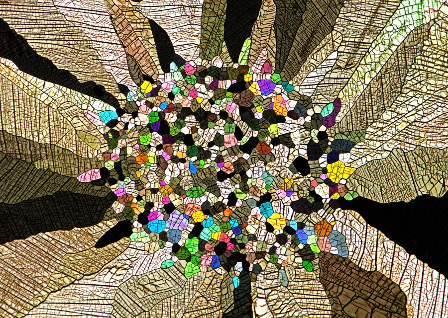
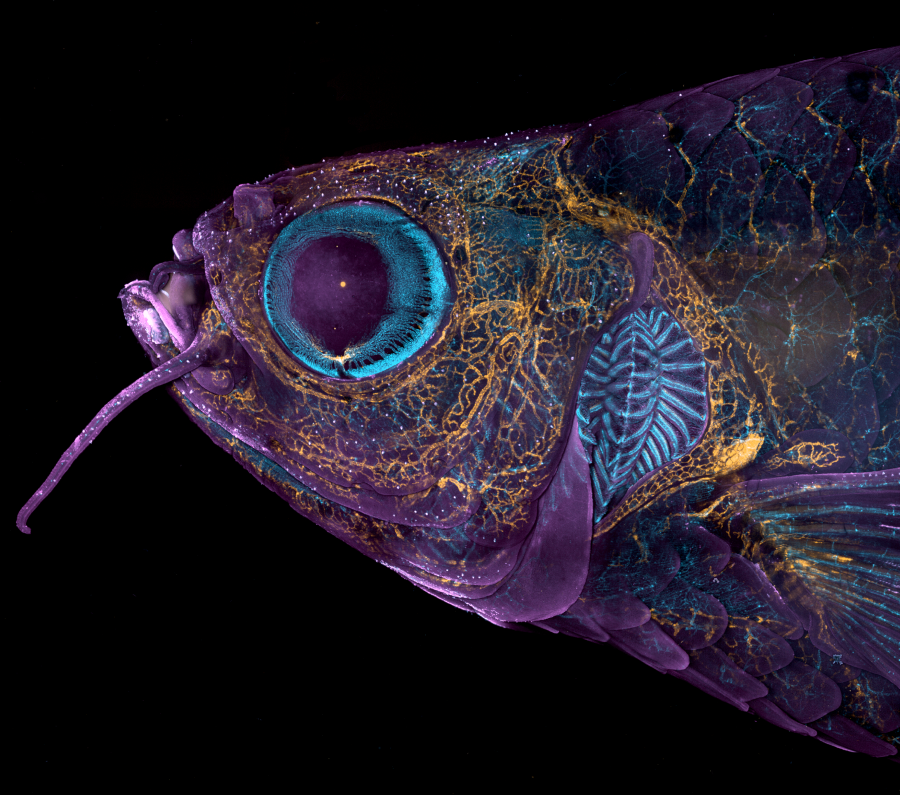
By way of a microscopic lens, the heat from an igniting matchstick – captured within one particular-8-thousandth of a next – is palpable. A mosaic of caffeine crystals could be mistaken for a get the job done of abstract artwork. And a massive, venomous fang plunging into body basically belongs to a 13-centimeter tarantula.

This calendar year marks Nikon’s 49th Little Earth Photomicrography level of competition, which acknowledges excellence in pictures through the microscope. Past their aesthetic attraction, these charming microscopic photos maintain the vital to advancing vital scientific analysis.

For occasion, this year’s 1st-spot winner is a colorful picture of a rodent optic nerve head, developed by Hassanain Qambari and Jayden Dickson. Qambari has used visuals like this to research diabetic retinopathy for two a long time.

He famous that Nikon’s opposition is an crucial opportunity to showcase scientific achievement. “All the photographs introduced in the opposition signify the splendor and artistic aspect of science, which may well usually get disregarded,” Qambari reported in a Nikon press launch.

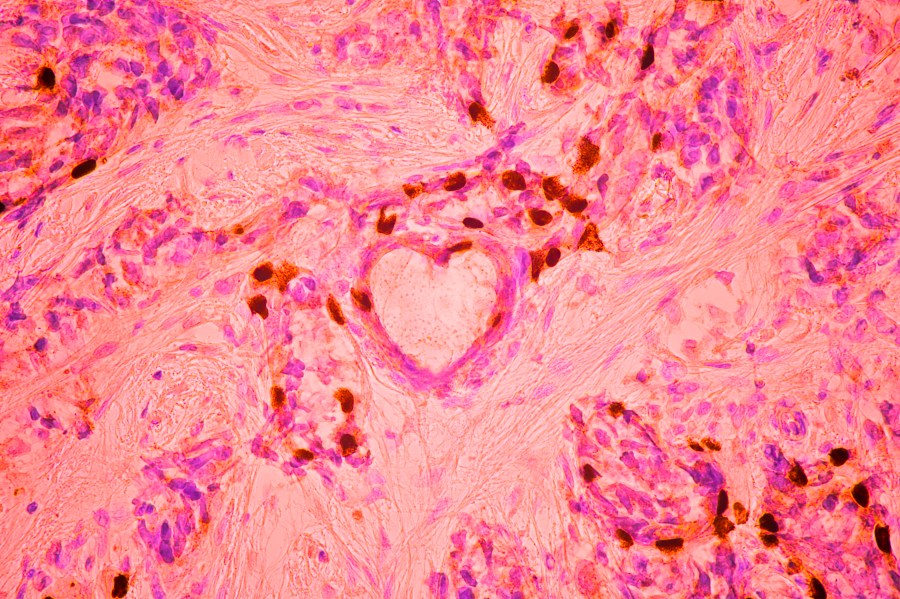
From the condition of a coronary heart nestled in a cluster of breast cancer cells to the translucent head of a zebrafish, the artistry of photomicrography is produced very clear by this year’s winners. In point, searching these photos feels like getting into a portal to yet another universe.

See the full listing of winners and honorable mentions right here.

Rodent optic nerve head exhibiting astrocytes (yellow), contractile proteins (crimson) and retinal vasculature (inexperienced). (By Hassanain Qambari and Jayden Dickson / Nikon Modest World)
Matchstick igniting by the friction floor of the box. (By Ole Bielfeldt / Nikon Modest Planet)
Breast most cancers cells. (By Malgorzata Lisowska / Nikon Little Environment)
Venomous fangs of a tiny tarantula. (By John-Oliver Dum / Nikon Modest Globe)
Auto-fluorescing defensive hairs masking the leaf surface of Eleagnus angustifolia exposed to UV gentle. (By Dr. David Maitland / Nikon Modest Earth)
Slime mould (Comatricha nigra) exhibiting capillitial fibers as a result of its translucent peridium. (By Timothy Boomer / Nikon Small Globe)
Mouse embryo. (By Dr. Grigorii Timin and Dr. Michel Milinkovitch / Nikon Tiny Earth)
Caffeine crystals. (By Stefan Eberhard / Nikon Tiny Globe)
Crystallized sugar syrup. (By Dr. Diego García / Nikon Tiny World)
“Cuckoo wasp” standing on a flower. (By Sherif Abdallah Ahmed / Nikon Tiny Entire world)
Sunflower pollen on an acupuncture needle. (By John-Oliver Dum / Nikon Modest Entire world)
Chinese moon moth (Actias ningpoana) wing scales. (By Yuan Ji / Nikon Tiny Globe)
Grownup transgenic zebrafish head exhibiting blood vessels (blue), lymphatic vessels (yellow), and the pores and skin and scales (magenta). (By Daniel Castranova and Dr. Brant Weinstein / Nikon Little Globe)


Source connection